Research Highlights
We have two goals: firstly, develop new optical techniques, and then use these techniques to solve some fundamental problems related to soft condensed matter such as single molecule manipulation and characterization in liquid, non-equilibrium dynamics at solid-liquid interface, water under confinement, etc.
A. Events near interfaces reported by new scanning probe microscopy
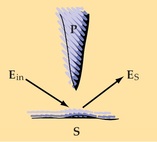
Tip-enhance Raman spectroscopy and fluorescence have experienced fast development in the past decade. In our group, we want to find new ways to enhance, probe, manipulate nanoparticles and molecules on surfaces. Recently, we start to build optical tweezer in combination with spectroscopic method so as to probe the interfaces of different materials. We hope to develop a method that is similar to tip-enhance SPM method, and also can be readily applied in water, with fast and easy 3D manipulate capability, physical topography mapping, and spectral signal enhancement. The combination of optical tweezers and laser spectroscopy can be used to study important mechanobiological properties of living orgnisms, such as those related to cancer development, cell motility, and mechanotransduction pathways. In this topic, we hope to use some well-defined polymer model systems to test our technique, and gradually move on to well-known cellular systems as model systems to show that new information (both physical and chemical) can be obtained.
B. Photothermal spectroscopy for sensing and imaging
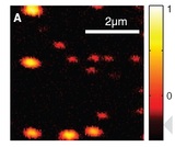
Metal nanoparticles with environment-specific surface plasmon resonance (SPR) can sense extremely small changes of the environment such as temperature, pH, ion concentration, and refractive index. Recently, light scattering from individual metal nanoparticle under dark field illumination scheme has been used to probe the SPR. However, light scattering method is restricted to particles larger than 30nm. Inherently, smaller particles are more sensitive to environmental change and can sense molecule binding events even at very low concentrations. In this project, we employ the sensitive photothermal effect (one-photon resonance and two-photon resonance) of gold and silver nanoparticles to probe interactions of organics molecules, ions, and biological species with metal particle surfaces. We are also developing gas sensing method based on photothermal imaging technique.
C. Spectroscopy of water under extreme physical conditions
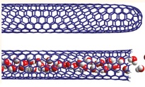
When water is confined in small space, the physical and chemical properties change as a consequence. For example, the flow of water through a carbon nanotube (CNT) is nearly frictionless and ballistic. Thin water film is shown to resist pH change and exhibit different buffer action. Although there have been man theoretical studies, there is very little spectroscopic evidence for such drastic changes. In these studies, we use a combination of infrared spectroscopy, fluorescence spectroscopy, and second harmonic generation (SHG) to probe the water molecules as well as water-solvated species confined in extremely small space. One typical model system that we currently investigate is the so called "black soap film" with a thickness of 10-100nm.
D. Submerged Microsphere Optical Nanoscopy (SMON)
The optical super-resolution imaging competition has reached an exciting length scale of 10nm during the past years. However, need for special phosphors, complicated labeling method, expensive laser equipment are some of the drawbacks of current fluorescence-based super-resolution techniques. In recent years, a simple and less-expensive method based on microsphere near-field imaging has emerged and started to show promising results.
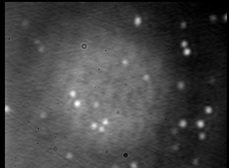
In our group, we are developing wide-field Microsphere Enabled Raman Super-resolution Imaging (MERSI) for imaging chemistry-relevant surface nanostructures. At the same time, we use microphere for direct observation and sizing of fluorescent and non-fluorescent nanoparticles, and direct mapping of plasmonic modes of individual and interacting NPs. (Image: 60nm GNPs under microsphere with white light illumination)
The optical super-resolution imaging competition has reached an exciting length scale of 10nm during the past years. However, need for special phosphors, complicated labeling method, expensive laser equipment are some of the drawbacks of current fluorescence-based super-resolution techniques. In recent years, a simple and less-expensive method based on microsphere near-field imaging has emerged and started to show promising results.
In our group, we are developing wide-field Microsphere Enabled Raman Super-resolution Imaging (MERSI) for imaging chemistry-relevant surface nanostructures. At the same time, we use microphere for direct observation and sizing of fluorescent and non-fluorescent nanoparticles, and direct mapping of plasmonic modes of individual and interacting NPs. (Image: 60nm GNPs under microsphere with white light illumination)
